This article delves into the world of Atopic Dermatitis, a common chronic skin condition characterized by inflammation, itching, and redness. By analyzing atopic dermatitis pictures, we can better understand the visual manifestations of this condition. These images assist healthcare professionals and patients in identifying and managing symptoms effectively, leveraging insights from visual evidence to aid both diagnosis and treatment planning.
Atopic dermatitis, commonly known as eczema, is a chronic inflammatory skin condition that afflicts millions globally, characterized by episodes of itchy, inflamed skin. The condition often begins in childhood, and although some children may outgrow it, many individuals experience persistent symptoms throughout their lives. Understanding and diagnosing this condition involves a significant focus on visual symptoms, making the study of atopic dermatitis pictures pivotal in its management and treatment. The term "atopic" refers to a group of conditions that include asthma and hay fever, indicating a hereditary tendency to develop allergic diseases. Consequently, atopic dermatitis is associated with a complex interplay of genetic, environmental, and immunological factors.
Images of atopic dermatitis are crucial in the diagnostic process. These pictures display various stages and severities of the condition, providing healthcare professionals and dermatologists with a visual reference to better understand the skin’s appearance during flare-ups. Recognizing these characteristics can aid in distinguishing atopic dermatitis from other skin conditions such as psoriasis or contact dermatitis. The use of visual references enhances clinicians' diagnostic accuracy and empowers patients to articulate their experiences better, leading to more informed treatment decisions.
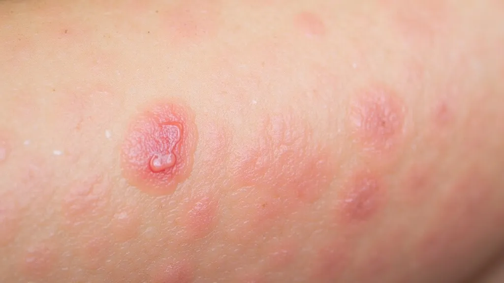
Images typically showcase areas of the skin affected by redness, swelling, and occasionally, oozing or crusting. Chronic cases may reveal lichenification, a leathery appearance due to prolonged scratching, and often show areas of hyperpigmentation afterward. The distribution of these lesions—often on the face, arms, and legs—offers clues to the diagnosis, as certain patterns can indicate the chronicity of the condition or potential triggers. Additional features that may appear in various stages include small, raised bumps that may leak fluid when scratched and crusting sore patches. A comprehensive gallery of atopic dermatitis pictures serves as a valuable resource for both patients and healthcare providers, assisting in distinguishing this skin ailment from others and providing insight into the condition's natural history.
Dermatologists rely on a detailed examination of these images to assess the disease’s progression and the patient’s response to treatment. Such visual documentation enables specialists to tailor therapy plans to individual patient needs more effectively, as treatment may require personalized interventions based on the patient's unique presentation of the disease. Moreover, these pictures serve educational purposes, helping patients recognize early symptoms and prompting them to seek timely medical advice. A consistent visual record of the condition aids in evaluating the efficacy of treatment regimens over time, ensuring that adjustments can be made when necessary. Additionally, sharing these images during consultations can facilitate discussions about treatment options, improving patient engagement and adherence to therapeutic protocols.
| Severity Level | Common Features |
|---|---|
| Mild | Minor itching, small patches of dry skin, occasional redness. |
| Moderate | Persistent itching, larger areas involved, noticeable redness, and scaling. |
| Severe | Intense itching, extensive redness, cracked skin that may bleed, and oozing lesions. |
Recent advancements in imaging technology have significantly improved the capability to capture high-resolution atopic dermatitis pictures, allowing for detailed analysis of skin textures and colors. These technologies include dermatoscopic imaging, which enhances the visibility of skin features, and 3D imaging systems that provide comprehensive assessments of skin structures. This technological evolution aids in both clinical settings and academic research, offering a clearer understanding of the condition’s development and persistence. The increased resolution allows clinicians to discern subtle changes in skin texture or color that may be overlooked in standard photography.
Moreover, artificial intelligence (AI) and machine learning algorithms are being integrated into dermatological practices to analyze images more effectively. These tools can assist in identifying patterns and predicting flare-ups based on historical data captured through imaging, leading to more proactive management strategies for patients. Teledermatology has also gained traction, allowing patients to submit images of their condition for remote consultation, which is particularly beneficial for those in rural or underserved areas. This accessibility removes traditional barriers to care and facilitates earlier interventions, ultimately leading to better outcomes for individuals suffering from atopic dermatitis.
Genetic factors, environmental triggers, and immune system dysregulation play significant roles in the etiology of atopic dermatitis. Individuals with a family history of atopic diseases are more susceptible, suggesting a genetic predisposition. Environmental factors such as allergens, irritants, and changes in weather conditions can exacerbate the condition, leading to flare-ups.
These images help dermatologists analyze and compare the visual characteristics of the skin, guiding them in confirming a diagnosis and differentiating it from other similar skin disorders. Images serve as both a diagnostic and monitoring tool, facilitating the tracking of changes over time in response to treatment protocols.
Yes, these images often show the extent of skin inflammation, which can serve as an indicator of flare-up severity. Dermatologists may classify severity levels based on clinical evaluation of images and correlate findings with patient-reported symptoms.
Common triggers include specific foods (such as nuts or dairy), environmental allergens (like pollen or pet dander), irritants (such as soaps, detergents, or fabrics), stress, and changes in temperature or humidity. Understanding personal triggers is crucial for individuals managing their condition, as minimizing exposure can help reduce flare-ups.
No, atopic dermatitis is not contagious. It is an intrinsic condition that arises due to a combination of genetic predisposition and environmental interactions. Individuals cannot contract atopic dermatitis from another person, although they might share the same environmental triggers that can provoke symptoms.
Management strategies include the regular use of moisturizers, topical corticosteroids, and calcineurin inhibitors to reduce inflammation and itch. Systemic medications and biologics may be necessary for severe cases resistant to standard treatments. Phototherapy is also an option for some individuals. Education about skincare routines, lifestyle modifications, and avoidance of known triggers is essential in effectively managing atopic dermatitis.
Yes, lifestyle modifications can significantly influence the severity and frequency of flare-ups. Emphasizing a consistent skincare regimen, managing stress levels, maintaining a balanced diet, and adopting protective measures against allergens can collectively contribute to improved skin health and reduced symptoms.
In the realm of dermatology, atopic dermatitis images are indispensable tools for diagnosis, patient education, and treatment tracking. They offer a visual narrative that can guide healthcare decisions and provide patients reassurance and clarity through visual understanding of their condition. As research continues to evolve, incorporating advanced imaging techniques and AI-driven analyses will refine the diagnostic and therapeutic pathways for atopic dermatitis, enhancing patient outcomes and quality of life. The journey towards understanding atopic dermatitis is ongoing, and expanding visual resources plays a fundamental role in this process, bridging the gap between clinician expertise and patient experience.
Explore the Tranquil Bliss of Idyllic Rural Retreats

Ultimate Countdown: The 20 Very Legendary Gaming Consoles Ever!

Understanding Halpin and its Influence

Affordable Full Mouth Dental Implants Near You
Discovering Springdale Estates

Illinois Dentatrust: Comprehensive Overview
Embark on Effortless Adventures: Unveiling the Top in Adventures Made Easy Outdoor Equipment

Unveiling Ossur Valves: Innovation in Prosthetics

Unlock the Full Potential of Your RAM 1500: Master the Art of Efficient Towing!

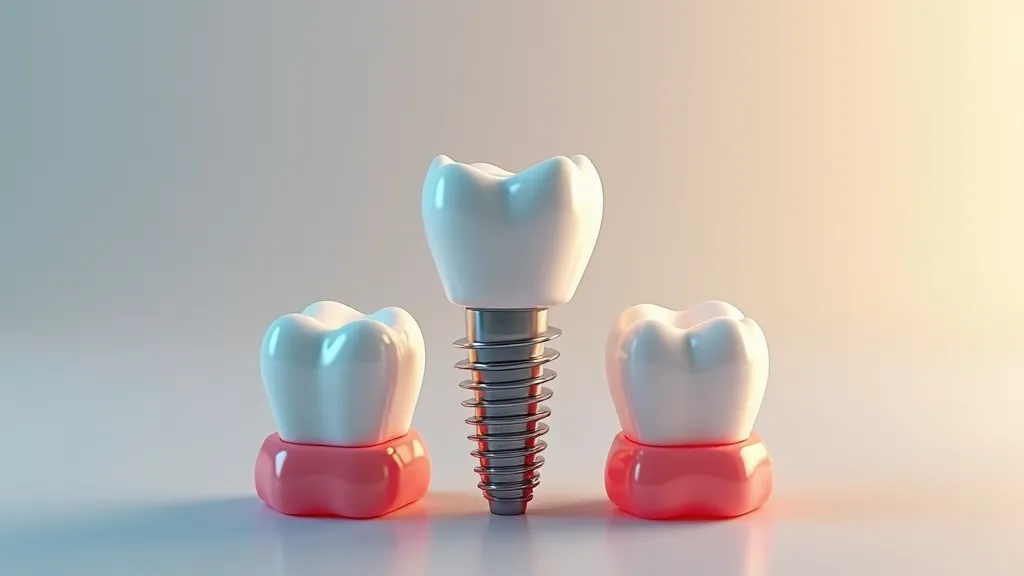
Revolutionizing Smiles: The Breakthrough Innovations in Dental Implants Changing Oral Health Care Forever
In the realm of dental medicine, the introduction of implant technology has dramatically transformed how professionals manage the absence of teeth. Providing more than just a stop-gap, these innovations have significantly influenced oral healthcare practices, offering alternatives that surpass older treatments like dentures and bridges, which often came with issues such as bone deterioration and persistent discomfort. By delving into the progression of dental implant advancements, we gain insight into their substantial contribution to the health and well-being of individuals with tooth loss.

Unveiling the Top Dental Implant Options for Seniors: Transform Your Smile with the Ultimate Guide to Restored Radiance
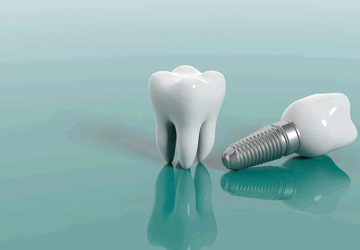
The significance of oral health cannot be understated for the elderly population as they are commonly afflicted with tooth loss due to a multitude of factors, including decay and gum disease. Dental implants have risen to prominence as a prime method of teeth replacement, acclaimed for their long-lasting nature, functional prowess, and striking similarity to our own teeth. This discussion delves deeply into the realm of dental implants to demonstrate why they are considered the optimal choice for aged individuals aiming to enhance their smiles and overall life quality.

Master the Art of Flossing for a Stunning Smile and Healthy Gums
Engaging in the routine activity of flossing is a key element in warding off periodontal diseases and sustaining prime dental hygiene. Despite its seeming simplicity, a great number of people are unaware of how to floss in the very efficacious manner. This discussion aims to demystify the optimal flossing practices and provide useful advice to enhance the benefits of your daily flossing regimen.

Discover the Key to a Dazzling Smile: Your Ultimate Handbook for Selecting the Ideal Tooth Replacement Option
Choosing to address the issue of absent dentition involves a thoughtful process and deliberate choices. The journey encompasses considerations ranging from visual appeal to practical use and financial implications, all contributing to the determination of the very appropriate form of dental restoration. The path, strewn with ample data and a variety of choices including implants, dentures, and bridges, may appear intimidating. This detailed handbook is crafted to shed light on crucial elements, assisting you in arriving at a well-considered and personalized decision based on your individual needs and condition.

The Gastric Sleeve Procedure Unveiled
Gastric sleeve surgery, also known as sleeve gastrectomy, is a weight-loss procedure where a significant portion of the stomach is removed, resulting in a tube-like structure. This procedure decreases the size of the stomach, helping patients feel fuller faster and aiding in sustainable weight loss. It is essential for individuals considering this surgery to understand its implications, benefits, and potential risks.

Guiding Your Family Through the Conversation: Navigating a Lung Cancer Diagnosis Together
Receiving a lung cancer diagnosis is not only a medical shock but an emotional one as well, particularly when it comes to telling your family. These heart-to-heart talks are laden with feelings and pose difficulties for everyone involved. To navigate these discussions about a lung cancer diagnosis with your loved ones, consider the following strategies.